lymphoma thyroid cancer ultrasound colors
The median age of patients was 56 2564 years. It is more common in women with an MF ratio of 125 range 116-31 2.
A thin hypoechoic capsule arrow is noted peripherally.

. Thyroid cancer presenting as a hot thyroid nodule. Staging is a tool your doctor uses to classify characteristics about your malignant thyroid tumor. Papillary thyroid cancer as is the case with follicular thyroid cancer typically occurs in the middle-aged with a peak incidence in the 3 rd and 4 th decades.
After thyroid cancer is diagnosed it is staged. 1 Although rare primary thyroid lymphoma should always be considered in the differential diagnosis of thyroid nodules or goiter. Distinguishing normal from malignant lymph nodes is critical for accurate staging prognosis and determination of optimal therapeutic options.
Ultrasound of thyroid cancer. Ad FDA Approved Differentiated Thyroid Cancer Treatment. 2D black and white image in 1mm slice.
Clinical diagnosis of PTL may not be easily established based on imaging studies as the imaging features of PTL are similar to those of lymphocytic thyroiditis and primary thyroid. Staging the tumor helps your doctor determine the best treatment for your thyroid cancer. Primary manifestation of subclinical systemic disease.
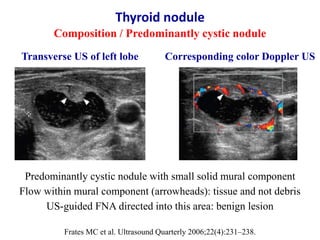
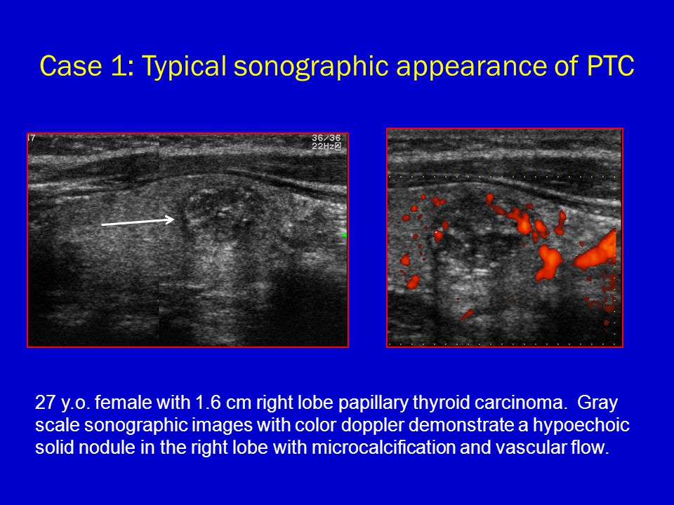
Ultrasound is the first-line imaging modality for assessment of thyroid nodules found on clinical examination or incidentally on another imaging modality. B The color Doppler ultrasound shows reduction of twisted blood flow signals in the primary thyroid lymphoma after three cycles of rituximab plus bendamustine. In A the two-part figure on the left shows a thyroid adenoma with a peripheral halo sign arrows in a nodule that is very well circumscribedThe companion color flow image shows a predominately peripheral pattern of flow suggestive of benign disease.
11 Rago T Vitti P Chiovato L et al. The staging system was developed by the American Joint Committee on Cancer AJCC and is called the TNM System. An early diagnosis of primary thyroid lymphoma can lead to timely treatment and a favorable prognosis.
It accounts for the majority 70 of all thyroid neoplasms and 85 of all thyroid cancers 24. The risk of cancer increased with the size of. Among 1156 newly diagnosed malignant lymphoma cases 8 were synchronous double primary lymphoma and thyroid cancer cases the crude incidence rate of synchronous thyroid cancer in lymphoma patients was 1384110 5 year.
Report of a case and review of the literature. This is called primary thyroid lymphoma to. Primary Thyroid Lymphoma.
Dr Anil T Ahuja Department of Diagnostic Radiology Organ Imaging The Chinese University of Hong Kong Prince of Wales Hospital 30-32 Ngan Shing Street Shatin New Territories Hong Kong SAR. Kim EH and Kim JY 2014 Aggressive primary thyroid lymphoma. On average 1 case of thyroid cancer was found for every 111 ultrasound exams performed.
Lymphoma is a cancer that develops in the lymphatic system the tissues and organs that produce store and carry white blood cells. Wang Z Fu B Xiao Y Liao J Xie P 2015 Primary thyroid lymphoma has different sonographic and color Doppler features compared to nodular goiter. Primary thyroid lymphoma diffuse large B-cell type in a 66-year-old man presenting with rapidly growing neck swelling.
Lymphoma usually occurs within lymph nodes but in rare cases it arises from lymphocytes that are present within the thyroid gland. This study was designed to investigate the sonographic features of PTL. Sonog-raphy shows the segmental type.
Objective Primary thyroid lymphoma PTL is an uncommon thyroid malignancy. Lower penetration higher resolution. Testicular lymphoma is an uncommon testicular malignancy.
Despite the rarity of PTL it is important to recognize PTL promptly because its management differs from that of all the other thyroid neoplasms. Primary site of extranodal disease primary testicular lymphoma secondary involvement of systemic disease. JTCC Specializes in Early Detection Risk Mitigation.
J Ultrasound Med 34. Transverse gray-scale ultrasound neck a shows a large well circumscribed oval shaped widthlength hyperechoic nodule in a thyroid lobe. A The gray-scale ultrasonography shows no significant decrease of the tumor size transverse diameters.
479 cm 473 cm 395 cm 301 cm. Stay Up-to-Date on Your Screenings. Microcalcifications were found in 38 of cancerous nodules and only in 5 of benign non-cancerous nodules.
2 However there are few reports about the. Color Doppler imaging shows a cen-. Ancillary features On grey scale ultrasound the presenceabsence of ancillary features such as matting of lymph nodes and adjacent soft tissue oedema should also be evaluated.
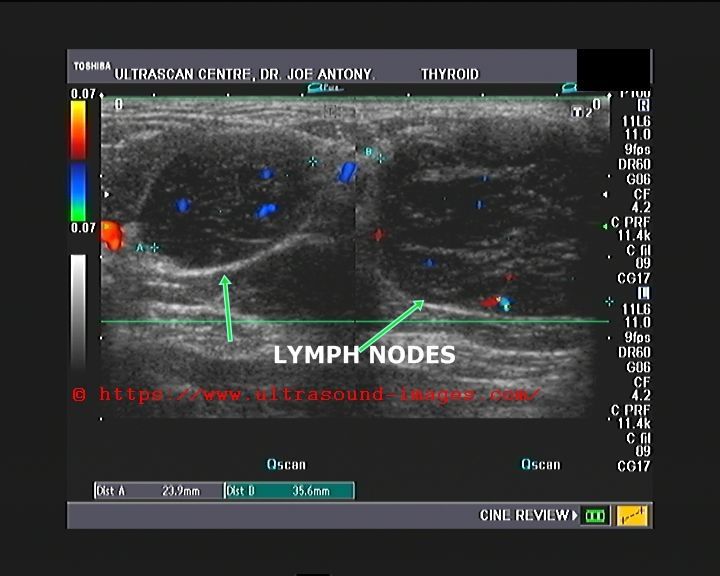
Transverse grey scale sonogram of a metastatic lymph node from papillary carcinoma of the thyroid arrows with echogenic punctate calcification arrowheads. Imaging features of two elderly patients. Thyroid nodules were found in 97 of patients with thyroid cancer and in 56 of without thyroid cancer.
Emitted waves are reflected back from the target material relative to the degree of the materials acoustic. Methods Twenty-seven pathologically confirmed PTLs were. Primary thyroid lymphoma PTL is a rare thyroid malignancy.
Various ultrasound findings in patients with a thyroid mass. Refining a differential diagnosis of thyroid nodules is usually a challenging clinical issue. Head and neck malignancies including squamous cell carcinoma lymphoma and thyroid cancer are a major cause of morbidity and mortality worldwide and frequently present with cervical lymphadenopathy.
This article is an overview of ultrasonographic features of thyroid nodules which are used to determine the need for biopsy with fine needle aspirationSpecific management guidelines from various. Lymphoma can involve the testes in three ways. Ad Check Our Screening Guidelines and Schedule Your Cancer Screenings Today.
The lesion has slight heterogeneous appearance due to presence of few tiny cystic spacesclefts. This article is concerned with primary testicular lymphoma. Role of conventional ultrasonography and color flow-doppler sonography in predicting malignancy in cold thyroid nodules.
Find Information for Patients. Wang et alSpecific Sonographic Features of Primary Thyroid Lymphoma 320 J Ultrasound Med 2015.
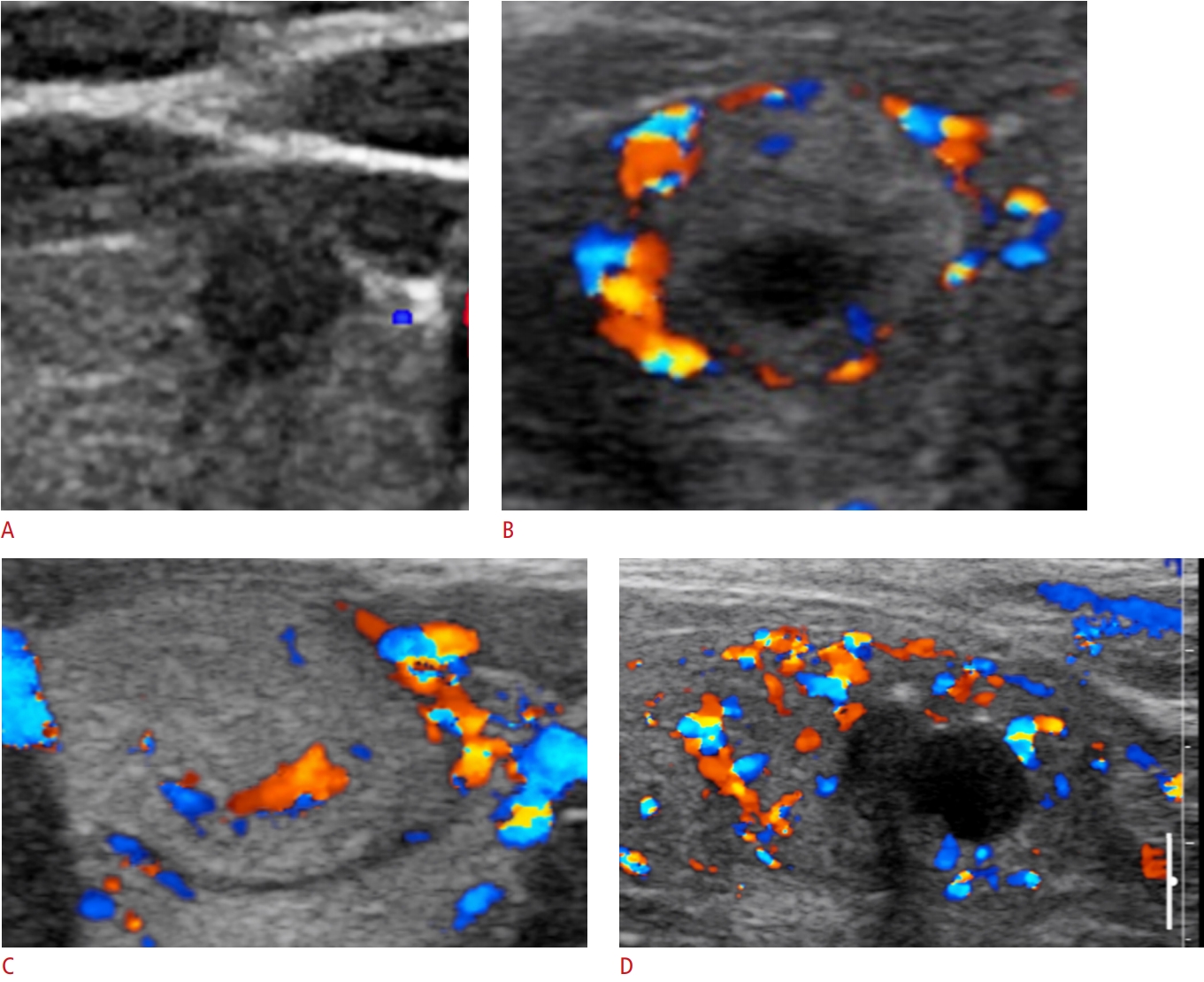
Usg Ultrasonography Ultrasonography 2288 5919 2288 5943 Korean Society Of Ultrasound In Medicine 10 14366 Usg 20072 Usg 20072 Review Article Clinical Applications Of Doppler Ultrasonography For Thyroid Disease Consensus Statement By The

Primary Thyroid Lymphoma Has Different Sonographic And Color Doppler Features Compared To Nodular Goiter Wang 2015 Journal Of Ultrasound In Medicine Wiley Online Library
Thyroid Nodules And Malignant Tumors Radiology Key

Evaluation Of Primary Thyroid Lymphoma By Ultrasonography Combined With Contrast Enhanced Ultrasonography A Pilot Study

Advances In The Management Of Thyroid Cancer Semantic Scholar

Application Of Color Doppler Ultrasound To Evaluate Chemotherapeutic Effect On Primary Thyroid Lymphoma

Color Doppler Patterns A Pattern 0 Normal Thyroid Vascularity B Download Scientific Diagram

Primary Thyroid Lymphoma Has Different Sonographic And Color Doppler Features Compared To Nodular Goiter Wang 2015 Journal Of Ultrasound In Medicine Wiley Online Library

Thyroid Lymphoma Nam 2012 Journal Of Ultrasound In Medicine Wiley Online Library
Other Malignancies In Thyroid Gland And Cervical Lymph Nodes Radiology Key
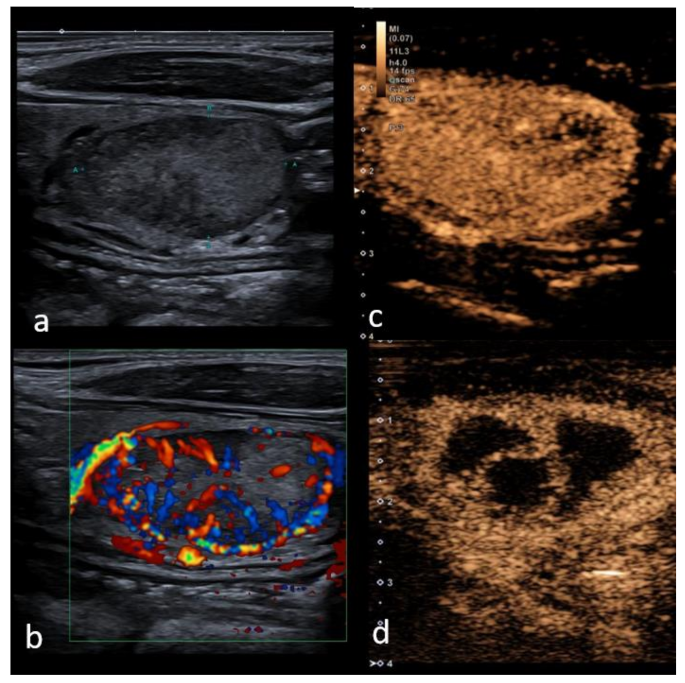
Cancers Free Full Text Performance Of Contrast Enhanced Ultrasound In Thyroid Nodules Review Of Current State And Future Perspectives Html
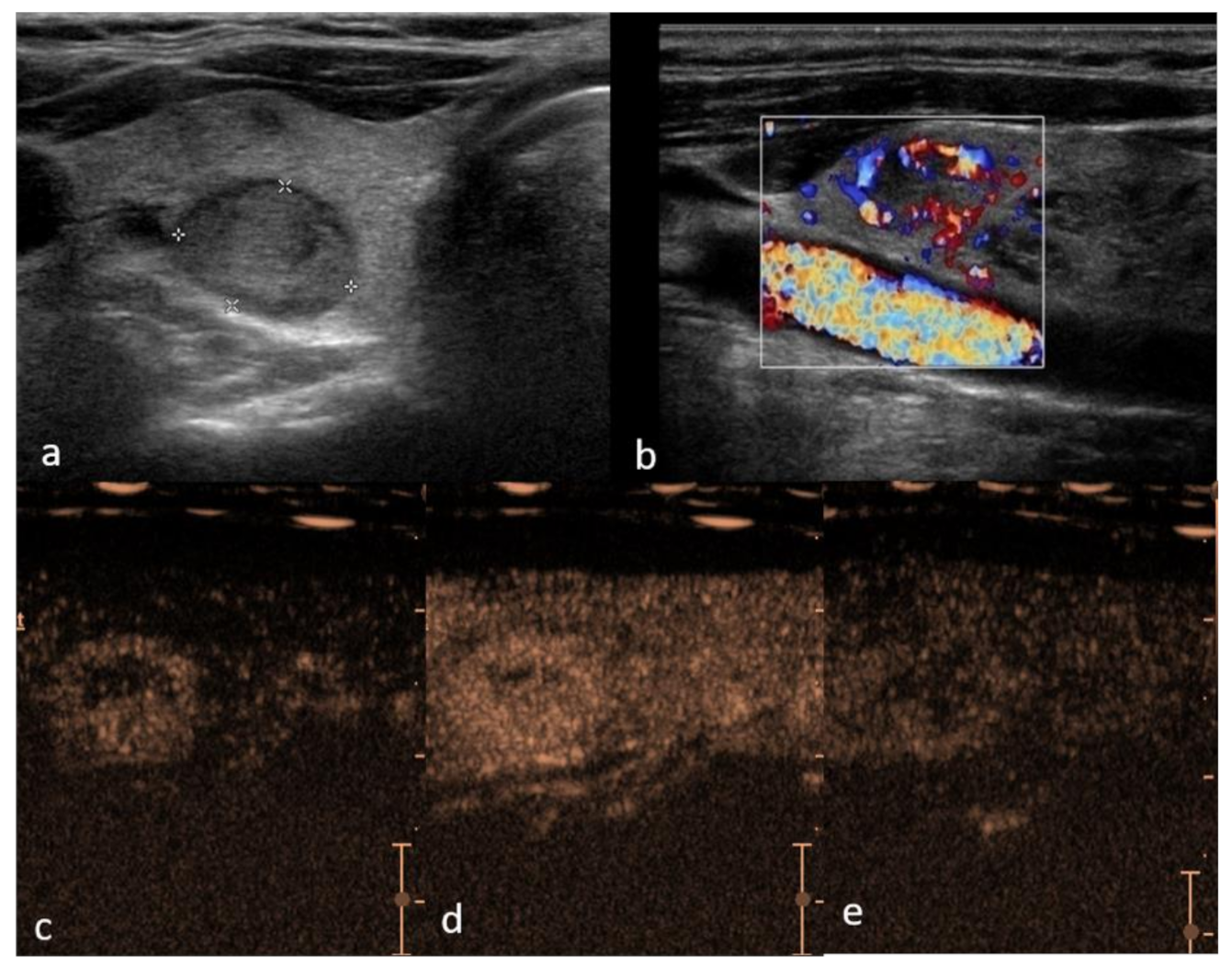
Cancers Free Full Text Performance Of Contrast Enhanced Ultrasound In Thyroid Nodules Review Of Current State And Future Perspectives Html

Primary Thyroid Lymphoma Has Different Sonographic And Color Doppler Features Compared To Nodular Goiter Wang 2015 Journal Of Ultrasound In Medicine Wiley Online Library

Normal Thyroid Gland A Gray Scale Ultrasound Transverse Scan Download Scientific Diagram

Figure 3 From Thyroid Ultrasound State Of The Art Part 2 Focal Thyroid Lesions Semantic Scholar
Imaging Of Differentiated Thyroid Cancer Ppt Video Online Download
A Gallery Of High Resolution Ultrasound Color Doppler 3d Images Lymphatic


